
Cervical Stenosis
Treatment in Noida
When your spinal canal narrows enough to squeeze the spinal cord, it stops being just a neck problem — your arms go weak, your hands lose precision, your walk becomes unsteady. Cervical stenosis is the most neurologically serious neck condition, and also the most treatable when caught before the cord sustains lasting damage. Noida's most advanced non-surgical decompression protocol is designed to protect your nervous system — before surgery becomes your only option.




Recognize the Warning Signs of Cervical Stenosis
Cervical Stenosis:
When Your Neck Starts Choking Your Nervous System
Of all conditions treated at 360 Neck Shoulder, cervical stenosis demands the highest clinical urgency. Not because it is the most painful — it often isn't. But because it is the only common neck condition that can silently compress the spinal cord and cause permanent neurological damage if left unaddressed. Understanding precisely what is happening inside a stenotic cervical spine — and what can still be done before surgery becomes unavoidable — is information every patient with this diagnosis deserves to have.
The cervical spinal canal is the bony tunnel formed by the vertebral bodies at the front and the laminae and ligaments at the back, through which the spinal cord descends from the brainstem. A healthy adult cervical canal typically measures 17–18 mm in its anteroposterior diameter. Clinical symptoms of cord compression begin when this diameter narrows below 13 mm — and become severe when it falls below 10 mm. The spinal cord, unlike peripheral nerves, has essentially no ability to regenerate once its axons are damaged by prolonged compression. This is the fundamental biology that makes early identification and treatment of cervical stenosis so clinically critical.
Cervical stenosis produces two distinct clinical syndromes depending on which neural structures are compressed. Foraminal stenosis — narrowing of the openings through which individual nerve roots exit the spinal canal — produces cervical radiculopathy: sharp, electric, burning pain and numbness radiating from the neck into one arm in a specific dermatomal pattern. The most commonly affected levels are C5-C6 (causing symptoms in the thumb, index finger, and outer forearm) and C6-C7 (causing symptoms in the middle fingers and outer forearm). Central canal stenosis — narrowing of the main spinal canal compressing the cord itself — produces cervical myelopathy: a more complex and serious syndrome affecting both arms and legs simultaneously, with progressive loss of hand coordination, gait disturbance, and in advanced cases, loss of sphincter control.
At 360 Neck Shoulder, we assess both components with precision — reviewing MRI findings for canal diameter measurements, cord signal changes (T2 hyperintensity indicating cord edema or damage), and foraminal narrowing patterns — before designing a treatment protocol that addresses both the structural decompression of the narrowed canal and the recovery of the compressed neural tissue.
Root Causes & Risk Factors
What Narrows the Cervical Canal:
The Four Structural Culprits
Cervical stenosis does not have a single cause — it is almost always a combination of structural factors that collectively reduce the available space for the spinal cord. Identifying which factors are dominant in each patient determines the specific treatment approach.
- Degenerative Disc and Osteophyte Complex: The most common cause of acquired cervical stenosis. As cervical discs desiccate and lose height with age, the disc bulges posteriorly into the canal. Simultaneously, the vertebral bodies develop osteophytes (bone spurs) along their posterior margins — these protrude directly into the spinal canal. Together, the posterior disc bulge and posterior osteophytes can reduce the available canal space by 4–6 mm at a single level. When multiple adjacent levels are affected simultaneously — the typical pattern in advanced spondylosis — the cumulative canal reduction becomes clinically significant even without severe stenosis at any single level.
- Ligamentum Flavum Hypertrophy: The ligamentum flavum — the elastic yellow ligament running along the posterior spinal canal — thickens progressively with age and degenerative loading. A healthy ligamentum flavum is 1.5–2 mm thick; in advanced degeneration, it can reach 4–6 mm, adding its entire thickness to the posterior canal narrowing. This is particularly significant in extension — when the neck bends backward, the ligamentum flavum buckles further inward, transiently but dramatically reducing the posterior canal space. This explains why many stenosis patients find neck extension reproduces their worst symptoms, including arm electric pain and leg weakness.
- Congenital Narrow Canal: Some individuals are born with a constitutionally narrower cervical spinal canal — a developmental variant rather than a disease process. These patients have less "buffer space" before degenerative changes produce symptomatic compression. A patient with a congenital canal of 14 mm who develops even modest spondylotic change — reducing the functional canal by 4 mm — will reach symptomatic compression thresholds far earlier than a patient with a 17 mm congenital canal undergoing the same degenerative changes. This is why some patients in their 40s develop severe myelopathy while others in their 70s with more degeneration remain asymptomatic.
- Ossification of the Posterior Longitudinal Ligament (OPLL): A condition particularly prevalent in patients of East and South Asian ancestry, OPLL involves calcification and bone formation within the posterior longitudinal ligament — the thick band running along the back surface of the vertebral bodies. As the ligament ossifies, it effectively becomes a bony ridge intruding directly into the spinal canal. OPLL can produce rapidly progressive and severe myelopathy that requires careful assessment to determine the role of physiotherapy versus neurosurgical intervention. Its higher prevalence in Indian and East Asian patients makes it an important diagnostic consideration in Noida's patient population.
Robotic Decompression — Precision Canal Opening Without Risk
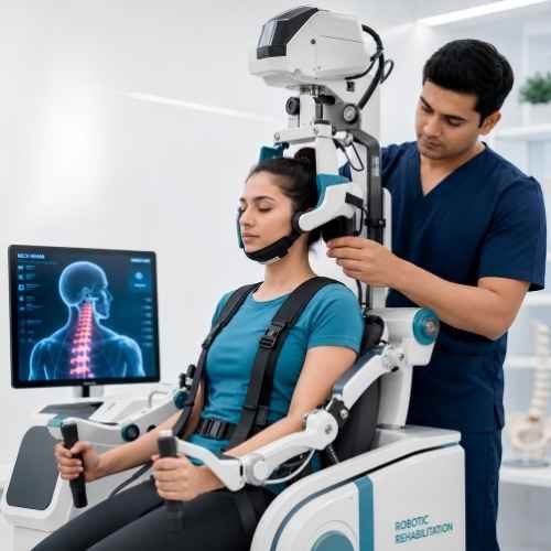
In cervical stenosis, forceful manual manipulation is strictly contraindicated — it risks worsening cord compression. Our robotic decompression system applies computer-controlled, real-time force-monitored traction calibrated to gently open the narrowed canal space. The system automatically limits forces if paraspinal muscle guarding is detected, making it uniquely safe for myelopathic patients where conventional manual therapy carries significant risk.
Decompression
Our Neurologically-Safe
Three-Stage Stenosis Rehabilitation
Cervical stenosis rehabilitation requires a fundamentally different philosophy from other neck conditions: first, do no harm. The proximity of the spinal cord to every intervention means that the treatment protocol must be as neurologically safe as it is therapeutically effective. Our three-stage approach is specifically designed around this imperative — progressing from the safest decompression modalities first, to progressive neurological recovery, to long-term canal protection.
Stage 1 — Controlled Canal Decompression & Neural Inflammation Reduction (Sessions 1–8)
The opening phase focuses on two simultaneous goals: mechanically increasing the available space within the narrowed canal and reducing the neurogenic inflammation that is amplifying the patient's symptoms beyond what the structural narrowing alone would produce. Robotic-controlled cervical traction is initiated at very conservative forces — beginning at 5–7 kg — and progressed carefully based on the patient's neurological response after each session. This is fundamentally different from traction protocols used in spondylosis: forces are lower, session duration is shorter, and the clinical team monitors neurological symptom response in real time during each session. Any increase in arm or leg symptoms triggers immediate force reduction and technique modification. TECAR therapy in Capacitive mode is applied to the paraspinal tissues and neural pathways — reducing the neurogenic inflammation component of the patient's symptoms and improving microcirculation to the compressed neural tissue. Most patients experience measurable improvement in arm tingling and numbness within 5–8 sessions as the combined decompression and anti-inflammatory effect takes hold.
Stage 2 — Neurological Recovery & Functional Rehabilitation (Sessions 8–18)
With the acute compression phase relieved, Stage 2 focuses on recovering the neurological function that was impaired by cord or nerve root compression — and this distinguishes cervical stenosis rehabilitation from other neck conditions. Patients with any myelopathic features — gait disturbance, hand clumsiness, proprioceptive loss — receive targeted neurological rehabilitation: balance board training for proprioceptive recovery, fine motor hand exercises for corticospinal tract reactivation, and gait retraining if coordination is affected. This stage also introduces progressive deep cervical flexor strengthening — in positions that do not compromise the canal — to begin building the muscular support system that will protect the canal long-term. Robotic range-of-motion training restores flexion and lateral mobility while maintaining safe limits on cervical extension, which remains restricted throughout Stage 2 due to the ligamentum flavum buckling risk.
Stage 3 — Canal Protection, Posture Correction & Long-Term Maintenance (Sessions 18–24)
The defining goal of Stage 3 is ensuring the stenosis does not progress further — and that the patient is equipped with the knowledge and physical capacity to protect their own cervical canal for life. The canal itself cannot be structurally widened by physiotherapy — the bone spurs and thickened ligaments remain. What changes is the dynamic component: the muscular and postural environment in which the canal exists. A cervical spine with strong deep stabilizers, corrected lordosis, and ergonomically optimized posture places significantly less dynamic compressive load on the canal than a poorly stabilized, kyphotic spine. Comprehensive ergonomic assessment — workstation, sleeping position, device use — is completed. A home exercise programme for lifelong maintenance is established and taught. Patients receive explicit guidance on the neurological warning signs that should trigger immediate re-assessment: new or rapidly progressing weakness, gait changes, or any bladder/bowel symptoms. For patients with moderate to severe structural stenosis, we maintain a quarterly monitoring protocol and provide clear surgical referral thresholds — so that if surgery does eventually become necessary, it is pursued proactively, not in crisis.
Patient Questions