
Cervical Spondylolysis
Treatment in Noida
A cracked or defective pars interarticularis — the tiny bony bridge holding your cervical vertebra together — is one of the most under-diagnosed causes of deep posterior neck pain in young adults and athletes. Most patients spend months being treated for the wrong condition. Noida's most precise diagnosis and targeted bone-healing rehabilitation is here.




Recognize the Warning Signs of Cervical Spondylolysis
Cervical Spondylolysis:
The Neck Fracture Athletes Don't Know They Have
Of the three "cervical spondy" conditions — spondylosis, spondylitis, and spondylolysis — the last one is the least known yet critically important to diagnose correctly. Cervical spondylolysis affects a completely different structure than the other two, occurs in a distinctly different population, and demands an entirely different treatment approach. Treating it as spondylosis — with generic traction and mobilization — can actually worsen the structural instability and delay recovery by months.
To understand cervical spondylolysis, picture the architecture of a single cervical vertebra. Each vertebra has a large body at the front and two pairs of articular processes (joints) at the back — the superior facets connecting to the vertebra above, and the inferior facets connecting to the vertebra below. The pars interarticularis is the narrow bony column that bridges these two sets of facet joints on each side. It is structurally elegant — but mechanically vulnerable. This slender bony bridge must resist enormous rotational and extension-compression forces every time the neck moves, and it is the pars that fails first under extreme or repetitive athletic loading.
Spondylolysis specifically means a defect, crack, or stress fracture in this pars interarticularis. Unlike cervical spondylosis (disc and facet joint degeneration) or cervical spondylitis (joint inflammation), spondylolysis is a structural break in the bone itself. It can occur in two ways: as an acute stress fracture from a single traumatic event (a collision, a fall during gymnastics, a wrestling takedown), or as a fatigue fracture from cyclic repetitive loading — the same mechanism that causes shin splints or metatarsal stress fractures in runners, but occurring in the vertebral pars.
In a third category of patients, the defect is congenital — the pars never fully formed during fetal bone development, leaving a structural gap that may remain painless for years until a sports injury or acute loading event triggers symptomatic instability. What is common to all three types is the characteristic clinical pattern: deep posterior neck pain that worsens specifically with neck extension and rotation, and partially relieves with neck flexion — the exact opposite of the mechanical pain pattern seen in cervical spondylosis.
Root Causes & Risk Factors
Who Gets Cervical Spondylolysis
and Why the Pars Fractures
Cervical spondylolysis is primarily a condition of young, active individuals — athletes, gym enthusiasts, and young adults with specific sport exposures. Understanding the mechanisms helps prevent recurrence after rehabilitation.
- Gymnastics and Acrobatic Sports: The highest-risk activity for cervical spondylolysis. Back walkovers, back handsprings, and aerial dismounts require repeated full cervical extension under axial loading — placing extreme compressive and shear forces directly on the pars interarticularis at the C5-C6 and C6-C7 levels. Elite gymnasts have a cervical spondylolysis prevalence several times higher than the general population. The condition is frequently bilateral in gymnasts — both left and right pars defective — increasing the risk of eventual spondylolisthesis.
- Contact Sports — Wrestling and Rugby: Spear tackling, headlocks, and impact loading of the hyperextended cervical spine in wrestling and rugby generate acute trauma at the pars. A single high-energy impact can create an acute pars fracture. Repetitive subconcussive impacts in practice sessions can create a fatigue fracture that progresses slowly and is often missed on early imaging because the pars cortex has not yet fully separated.
- Swimming — Butterfly and Backstroke: The butterfly stroke demands repeated full cervical extension with forceful head lift out of the water against resistance. Elite butterfly swimmers perform thousands of these repetitions per training season — creating a fatigue-loading pattern on the posterior cervical pars that can produce stress fractures even without any acute injury event. Backstroke swimmers experience a similar mechanism through the repeated rotational head-turn pattern at the entry phase.
- Overhead Throwing and Weightlifting: Javelin, cricket bowling, and overhead press movements in weightlifting create extension-compression loading at the cervical pars during the follow-through or lockout phases. Olympic lifters who perform repeated clean-and-jerk or snatch movements with suboptimal cervical control are particularly vulnerable to progressive pars stress fractures in the mid-cervical spine.
TECAR Resistive Mode — Bone Healing Acceleration
For cervical spondylolysis, TECAR's Resistive mode targets the relatively poorly vascularized pars interarticularis bone directly. The deep electromagnetic energy stimulates osteoblast (bone-forming cell) activity, dramatically increases local vascular supply to the fracture site, and creates a biologically optimized environment for pars union — achieving non-invasively what bone stimulators attempt electrically, without implants or injections.
Healing
Our Three-Stage
Pars Healing & Return-to-Sport Protocol
The fundamental principle of cervical spondylolysis rehabilitation is sequenced: the pars defect must first be given the biological conditions to heal, then protected by muscular stabilization as it heals, and finally prepared for the sport-specific loading demands that caused the injury in the first place. Skipping any stage risks either non-union of the fracture or re-injury on return to sport.
Stage 1 — Load Management & TECAR Bone-Healing Therapy (Weeks 1–4)
The acute phase demands one non-negotiable intervention: removal of the repetitive extension-rotation loading that caused the pars fracture. This does not mean complete bed rest — but it does mean immediate sport modification and elimination of all cervical extension-dominant activities. Simultaneously, TECAR therapy in Resistive mode is applied directly over the identified pars defect level — typically guided by CT scan findings showing the fracture site. The Resistive mode energy penetrates the superficial cervical muscles to reach the posterior vertebral elements where the pars is located, stimulating the reparative biological cascade: osteoblast proliferation, angiogenesis (new blood vessel formation to the fracture site), and extracellular matrix deposition for callus formation. Patients typically report a reduction in baseline pain within 3–4 sessions as local inflammation at the fracture site resolves.
Stage 2 — Cervical Stabilization & Pars Protection (Weeks 4–10)
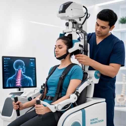
As healing progresses — confirmed clinically by reduction in extension pain and, where indicated, by follow-up SPECT or CT imaging — Stage 2 introduces progressive muscular stabilization of the cervical spine. The target muscles are the deep cervical flexors (longus colli, longus capitis) and the suboccipital extensors — working as an integrated system to provide dynamic control of cervical segmental motion, protecting the healing pars from excessive extension-rotation loads during daily activities. Critically, all exercises in this phase are performed in positions that avoid cervical extension — all strengthening is conducted in neutral and flexion-biased positions. Robotic-assisted range-of-motion training is introduced during late Stage 2 to restore pain-free flexion and lateral motion while continuing to respect the extension restriction. Progress through Stage 2 is individually paced based on symptom response and clinical milestones, not on a fixed calendar timeline.
Stage 3 — Sport-Specific Rehabilitation & Return to Activity (Weeks 10–20)
The most detailed and clinically demanding stage — and the one most responsible for preventing re-injury. Stage 3 progressively reintroduces sport-specific cervical loading in a controlled, monitored sequence. For gymnasts: extension loading is reintroduced in supported positions, then progressively loaded to full back walkover mechanics over 8–10 weeks. For wrestlers: resistance-band neck training in all planes, progressing to partner resistance, then controlled sparring before full contact return. For swimmers: water training is reintroduced with modified stroke technique, progressing to full butterfly training only after documented full cervical extension tolerance under load. Throughout Stage 3, TECAR maintenance sessions continue bi-weekly to support ongoing bone consolidation at the healed pars site. Return to full competitive sport is confirmed only when the athlete demonstrates: (1) full cervical extension under loading without pain; (2) deep cervical flexor strength symmetry; and (3) sport-specific movement patterns without compensatory guarding.
Patient Questions