
Cervical Spondylosis
Treatment in Noida
Cervical spondylosis is the single most common cause of neck pain in adults over 35 — and the most undertreated. Worn discs and bone spurs don't just cause neck pain; they compress nerves and, in advanced cases, the spinal cord itself. Noida's most advanced robotic decompression and TECAR protocol stops the degeneration cycle — permanently, without surgery.




Recognize the Warning Signs of Cervical Spondylosis
Cervical Spondylosis:
What Is Really Happening Inside Your Neck
Cervical spondylosis is the most common structural diagnosis in adult neck pain — and also one of the most misunderstood. Patients are told "your neck is degenerating" and handed a prescription for painkillers. But understanding precisely what is degenerating, why it is happening, and most importantly, what can be done about it — that is the difference between temporary relief and lasting recovery.
The cervical spine consists of 7 vertebrae (C1–C7) separated by intervertebral discs — gel-filled shock absorbers that maintain space between vertebrae and cushion spinal movement. In cervical spondylosis, these discs gradually lose their water content and height — a process called disc desiccation. As disc height decreases, the vertebral bodies above and below move closer together. The body responds by forming osteophytes (bone spurs) at the vertebral edges — its attempt to stabilize the degenerating segment. Simultaneously, the facet joints — the small paired joints at the back of each vertebra — develop osteoarthritis from the altered mechanical loading.
The clinical consequences become serious when these structural changes impinge on neural structures. Narrowed disc space combined with bone spurs can compress the cervical nerve roots as they exit through the foramina — causing the characteristic radiating arm pain, numbness, and weakness known as cervical radiculopathy. In more advanced cases, osteophytes can narrow the central spinal canal itself, compressing the spinal cord — a condition called cervical myelopathy, which requires urgent intervention.
At 360 Neck Shoulder Advanced Rehabilitation Centre, we treat the complete pathological chain — not just the symptom. Our protocol decompresses the narrowed disc space, reduces nerve root irritation, restores disc nutrition, and rebuilds the muscular support system that protects the cervical spine from further degeneration. The result is not just pain relief but genuinely slowed disease progression.
Root Causes & Risk Factors
Why Cervical Spondylosis Develops:
Beyond Just "Age-Related Wear"
While aging is the primary driver of cervical disc degeneration, the rate and severity of spondylosis are heavily determined by modifiable mechanical factors — particularly relevant for Noida's working population.
- Sustained 'Tech Neck' Compressive Loading: Every 10 degrees of forward head tilt increases the effective weight load on the cervical spine exponentially. At the 30–45 degree forward head position — standard for laptop and smartphone use — the cervical discs bear 18–27 kg of compressive force, compared to 5 kg in neutral posture. Over 8–10 hours of daily desk work across years, this sustained axial compression accelerates disc desiccation by impeding the osmotic fluid exchange that keeps discs hydrated and resilient. This is the single largest accelerator of early-onset cervical spondylosis in the 30–50 age group.
- Loss of Cervical Lordosis — The "Military Neck" Problem: The natural cervical spine has a gentle inward curve (lordosis) that distributes mechanical load evenly across disc surfaces. Prolonged forward head posture progressively flattens or even reverses this curve. A straight or kyphotic cervical spine concentrates all compressive load on the anterior disc margins — accelerating disc degeneration at specific levels (most commonly C5-C6 and C6-C7) while creating facet joint overloading at the posterior elements.
- Disc Herniation Precipitating Accelerated Degeneration: A herniated cervical disc — where the nucleus pulposus ruptures through the annulus fibrosus — is not merely an acute injury. The biochemical cascade triggered by disc herniation (inflammatory cytokine release, prostaglandin flooding) accelerates the degeneration of the affected disc and its adjacent levels. Herniation-triggered degeneration progresses significantly faster than natural age-related disc desiccation if not properly rehabilitated.
- Occupational and Genetic Risk Amplifiers: Beyond posture, certain occupational exposures dramatically accelerate spondylosis: sustained vibration (long-distance drivers, heavy equipment operators), repetitive overhead work, and heavy manual lifting with axial load. A family history of early-onset spondylosis is also clinically significant — genetic variation in disc collagen composition determines individual susceptibility to desiccation, independent of mechanical loading.
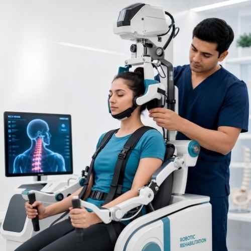
Robotic Cervical Decompression System
Our computer-controlled robotic traction system applies precise, reproducible decompressive forces calibrated to each patient's disc pathology level. Unlike manual traction, the robotic system adjusts in real-time to paraspinal muscle resistance — achieving true intervertebral disc distraction at C4-C5, C5-C6, and C6-C7 simultaneously, restoring disc space height and relieving nerve root compression with measurable clinical precision.
Decompression
Our Evidence-Based,
Three-Stage Decompression Protocol
Effective cervical spondylosis treatment must address three distinct pathological layers: the structural decompression of the narrowed disc space, the healing of the irritated nerve root, and the rebuilding of the deep muscular stabilization system that protects the disc from further mechanical stress. Treating only one or two of these layers is why most patients experience recurring pain despite treatment.
Stage 1 — Robotic Cervical Decompression & Nerve Root Decompression (Sessions 1–8)
The primary structural intervention. Our robotic decompression system applies computer-calibrated cervical traction forces — typically beginning at 8–10 kg and progressively increasing to 12–16 kg based on individual tissue response. This mechanically widens the narrowed intervertebral foramina, reducing direct pressure on the compressed nerve root. Simultaneously, the distraction of the disc space creates a negative intradiscal pressure — a vacuum effect that draws retracted disc material back toward the center of the disc, reducing the effective disc herniation or bulge. Patients typically experience arm pain and numbness relief within the first 4–6 sessions as nerve root decompression is achieved. TECAR therapy in Resistive mode is applied concurrently to the disc and facet joint levels to reduce concurrent inflammation and restore disc tissue nutrition through enhanced microcirculation.
Stage 2 — Segmental Mobilization & Facet Joint Rehabilitation (Sessions 8–16)
With acute nerve root compression relieved, Stage 2 addresses the facet joint osteoarthritis and capsular stiffness that restrict normal cervical movement and contribute to ongoing pain. Our precision manual mobilization techniques — combined with robotic-assisted range-of-motion training — restore the gliding mechanics of each individual cervical facet joint pair. This stage specifically targets the restoration of cervical lordosis — the natural inward curve that distributes mechanical load correctly across the disc surface. Restoring lordosis is critical: a cervical spine that regains its natural curve experiences significantly reduced compressive loading at the anterior disc margins, dramatically slowing further disc degeneration. TECAR's Capacitive mode is used during this phase for paraspinal muscle release and facet capsule healing.
Stage 3 — Deep Cervical Flexor Strengthening & Postural Correction (Sessions 16–24)
The definitive protection against recurrence. Research consistently demonstrates that patients with cervical spondylosis have severely weakened deep cervical flexor muscles — the longus colli and longus capitis — which are the primary active stabilizers of the cervical disc space. When these muscles are strong and coordinately active, they maintain the cervical spine in a loaded-but-protected position during work and daily activities. When they are weak — as they inevitably become in spondylosis — the superficial muscles (sternocleidomastoid, scalenes) overcompensate, creating chronic neck tension and abnormal compressive loading patterns that accelerate further degeneration. Our highly specific deep cervical flexor protocol — using real-time biofeedback — systematically retrains these muscles to provide 24/7 disc protection even during prolonged desk work. Combined with comprehensive ergonomic correction and home exercise programming, this stage is the patients' long-term insurance against relapse.
Patient Questions